Senior Distance Running Essentials Series
Chapter #5: Biomechanics of the Lower Extremity (May 2024)
Welcome to Chapter #5 of Senior Distance Running Essentials. If you have not read Chapter #4, which covers the principles of biomechanics, I suggest you read that now as it provides essential background for this chapter.
We are now going to examine what happens in our lower extremity when we run, starting with the hip and moving downward.
The Hip

The hip is the center of our body’s action. It provides a base for the upper body: our head, spine, chest, ribs, and arms. It also serves as the point of initiation for movement of our legs.
This figure shows the hip and upper leg from the front. Note the many attachment sites for muscles along the hip as well as cavities and depressions for insertion of bony structures or housing for muscles.

The best known of these cavities is the acetabulum, which accepts the head of the femur. This is commonly called our hip joint. Shown here, it is a ball-and-socket joint. The ball can rotate and spin in the socket, making it a fairly free-range joint, with movement in all three dimensional planes. In spite of this, the hip is quite stable due to the solid fit of bones and cartilage and the ligaments, tendons, and muscles that surround it.
The hip is a functional structure. As shown above, it is formed by the joining of three bones: the ilium, pubis, and ischium that are connected by immovable joints. Together, this configuration is known as our pelvis. The pelvis forms a ring, which protects everything inside of it and transfers weight from the upper to lower body.

Looking now at the rear view of the pelvis and upper leg, we see attachments for our posterior muscles, including our glutes and hamstring muscles, so critical for running.
As noted in Chapter #4, muscles have attachments at either end, called the origin or insertion. The origin usually grounds movement whereas the insertion is generally attached to the bone that is moving.

When we raise, or flex, our leg, the rectus femoris, which is one of our quadriceps whose origin is on the pelvis and the insertion on our tibia below the knee, provides the force to lift the leg. But this muscle has two functions: flexing our hip as shown below and which happens at the beginning of the gait cycle, and then extending our lower leg at the knee later in the cycle. As such, the hip is a base for movement in the lower extremity, even though it is positioned above our legs.

In the gait cycle, flexing our hip and lifting our leg allows us to move forward. We reverse this and prepare for the next stride as our leg extends behind us. Recall that muscles can only pull. Thus, muscles are paired with those usually on the opposite side of a limb to enable this forward movement.
There are six muscles directly involved in flexing the hip and five in extending it while other muscles play secondary roles. If any of these eleven muscles are stiff, relatively weak or strong, or strained causing pain, our gait will not be rhythmic and symmetrical. We know what that feels like – we simply don’t have our normal “bounce.” As we age, it is absolutely critical to maintain balances in strength and flexibility in these muscles. We will demonstrate various exercises you might employ in the strength training chapter.

Looking back at the hip joint where the head of the femur fits into the acetabulum, we can see how without that joint functioning as intended, we are going to have problems running. The wearing down of the hip joint is accelerated if there are muscle imbalances or structural deformities. Those who have run heavy mileage but shunned adequate recovery and strength training often have problems with this vital joint.
As has been noted, movement of our bones and therefore our bodies is made possible by muscle attachments. Below is a chart of the quadriceps muscles with their origins at the hip and femur and insertions below the knee, showing primary actions initiated by these muscles.
This is a whistle stop tour of the hip. Things that go wrong, if not corrected, can become chronic, which may lead to awkward walking and running. We all know people who have had one or both hips replaced. This often involves a new or partially new femur stem and head with replacement of the lining and concave surface of the acetabulum. And while medical technology continues to improve these replacement components, our first choice should be to support and maintain our original equipment.
| Muscle Name | Origin | Insertion | Primary Action(s) |
| Rectus Femoris | Anterior pelvis | TIBIA | Hip flexion, Knee extension |
| Vastus Intermedius | FEMUR | TIBIA | Knee extension |
| Vastus Medialis | FEMUR | TIBIA | Knee extension |
| Vastus Lateralis | FEMUR | TIBIA | Knee extension |
The Femur
As we move down the lower extremity toward our knee, we encounter the femur, the largest bone in our body.

While it looks straight, it actually bows slightly when under weight. This bowing enables the femur to bear more rather than less weight. At the distal end, just above the knee, the femur splits into two large segments, called condyles, helping to form the knee joint. This joint is the source of many running injuries, which is not surprising. When we run, jump, pick up a heavy object, or do a squat, our knee is right in the middle of those movements and is subject to a lot of force.
The Knee
Let’s look again at the picture of the knee from Chapter #4, which suggests the importance of ligaments and menisci in maintaining the integrity of the knee, and then another view on the right which shows more of the inter-workings of the knee.


These views show the intricate manner in which the parts of the knee must fit and work together. The cartilage absorbs shock and with sufficient synovial fluid (a synovial joint is encased in a fluid-filled capsule that provides lubrication) allows for smooth movement. Fluid is also a major ingredient of this cartilage.
Move it or lose it!
With age, we tend to “dry out,” often losing fluid content in our articular cartilage, which makes it less effective at shock absorption and friction reduction. However, we can mitigate this by understanding that cartilage, like many other tissues, stays strong with movement and pressure. A healthy knee that is moved and used in daily weight bearing challenges (i.e., walking, running, resistance training, etc.) is more likely to retain its cartilage as we age. This is a demonstration of Woolf’s Law, which we have previously considered and postulates that pressure stimulates growth.

Unlike many court and field sports, where there is a lot of twisting and turning resulting in torn ACLs, runners are typically moving forward in one direction. The greater issue with us is the repetitive nature of our sport. We all know people who have stopped running due to achy knees, often from worn cartilage, torn menisci, or patella pain, often called “runner’s knee.”
Runner’s Knee often stems from imbalances or weaknesses somewhere along the kinetic chain. For example, if the muscles surrounding the knee are not kept strong, tracking of the patella can be off, leading to pain in that area. I had a bout of patellar tendinitis after running my first marathon at age 32. I dealt with it off and on for a few years before discovering that regular leg extensions firmed up the quad muscles, which supported the kneecap and resolved the issue. I’ve been fortunate to avoid further knee problems since.
Let’s think a bit more about our quads. Have you ever wondered why we have four of them?
The reason follows their function and as with most everything in the body, form follows function.
| Muscle Name | Origin | Insertion | Primary Action(s) |
| Rectus Femoris | Anterior pelvis | TIBIA | Hip flexion, Knee extension |
| Vastus Intermedius | FEMUR | TIBIA | Knee extension |
| Vastus Medialis | FEMUR | TIBIA | Knee extension |
| Vastus Lateralis | FEMUR | TIBIA | Knee extension |
Above is the chart we looked at earlier. We see one of the quads attaches to the hip contributing to hip flexion. The other three attach to the femur. All four insert below the knee stretching across the patella, thus enabling knee extension. Do we really need all four? Well, yes! If we consider how the patella protects the inner knee, that it essentially floats, and that it is a rather odd shape, the four quads provide something of an envelope that serves to keep the patella in place as well as supplementing the overall support of the knee joint. It’s a package deal!
Assess problems early
So, how can we retain the integrity of our knees as we age? The first order of business is to not run through knee injuries. Find out why you are having a problem and address it early in the injury cycle. Experienced physical therapists can help assess imbalances. Also, be vigilant about finding suitable alternative forms of aerobic exercise, such as water running, cycling, elliptical, that use the knee joint but stress it less than running for some of your training load,
Number of running days
With age, or even well before you reach 60, it makes good sense to limit your number of running days per week, probably to no more than five, or if you run everyday alternate hard days with easier and shorter days. We will cover this in Part 2 of the series.

Like the hip, we’ve covered just a few basic aspects of the knee. It’s hard to overstate the importance of healthy knees, not only for running but daily living.
If we’re sitting in a concert and our knee is aching, it sure detracts from the performance! Remember too that proper function of our knee (and of all our joints) is a balancing act. We’ve looked at how our quads extend our knees and our hamstrings flex them. We need to keep ALL of these muscles strong and healthy for effective gait and peak performance.
Ankle/Foot
We now make it down to the ankle and foot, where the rubber meets the road! Or flesh if you run barefoot! The foot gets a lot of attention in the running world. When I worked in a running store in Boston, hardly anyone came in asking about their hips! While many did talk about their knees, the reason most came in was for shoes. Even after many hours of training and then experience on the floor, I was often baffled by the question “what is the best shoe for me?” What became apparent was there was no simple answer; that I needed to know what kind of running the person was doing: how much; how often; type of terrain, and what kind of injuries they had had. Of course, we watched them walk in bare feet, and then after trying on some shoes went outside and watched them run. I’d like to think we did an OK job in fitting most people.
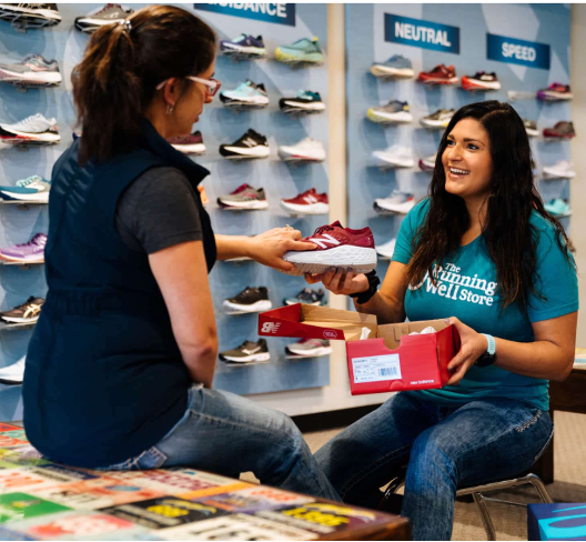
But it’s complicated. Some people are easy to fit, others not so. Ideally, you are able to find a specialty running store with knowledgeable staff who will work to find the right shoe or shoes for you.
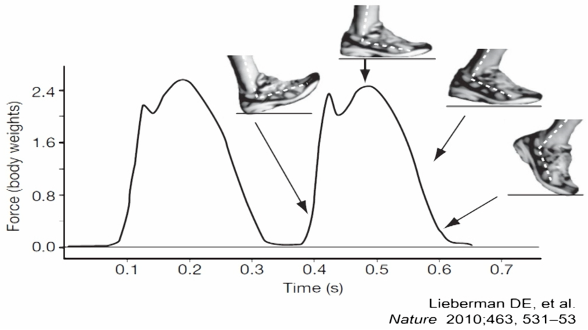
It’s hard to overstate the importance of the foot when we run! How we land on our foot and what happens as we move through the gait cycle is absolutely critical. Shown here is a graph illustrating the ground reaction forces experienced during the running cycle.
The puzzle, of course, is that it’s not just about the foot. Rather it’s the entire kinetic chain, which by now you may be tired of hearing about!
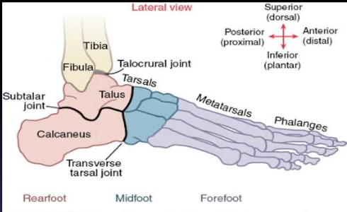
We have 26 bones & 33 joints in the ankle/foot. This is not surprising considering the range of terrain and surfaces the foot must navigate. This is enabled by a number of interlocking bony parts, as shown here.
All of these bones and joints are necessary because our foot plays 2 essential and opposing roles during our running gait cycle:
First, the foot is the initial shock absorber of impact when we make contact with the ground, and so it must be flexible and mobile to help absorb forces (that can be more than 3x your body weight!).
Second, the foot is the primary lever used during the push-off phase of running, and so it must be locked and rigid to help us transmit as much force as possible into the ground for a powerful push-off. The numerous bones and joints allow our feet to play both of these contradictory roles in a process so seamless we aren’t even aware it is happening!
The image below shows that the ankle is really a continuum of the lower leg. The tibia and fibula articulate with the talus to form the synovial talocrural joint, which is commonly called the ankle joint. The above image shows how bones forming the ankle joint are concave and convex, allowing for more stability. Which is a good thing when you consider almost all our weight, including two to three times our body weight when running, comes down on that joint!
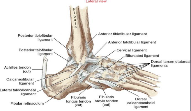
The other three components of our ankle and foot are the ligaments, tendons, and the muscles that provide increased stability and facilitate movement. When we say we sprained our ankle, what we really mean is we stretched (or tore!) some key ligaments beyond their allowable range. We can see that a number of ligaments the surround the ankle attach to both the leg and foot. They counterbalance each other, which is why when one is overstretched our stability is compromised, and the foot can’t function normally.
We don’t have to look too closely at this part of our body to marvel at how well the ankle and foot are designed and when working properly allow us to roll up onto our toes and then push off to run.
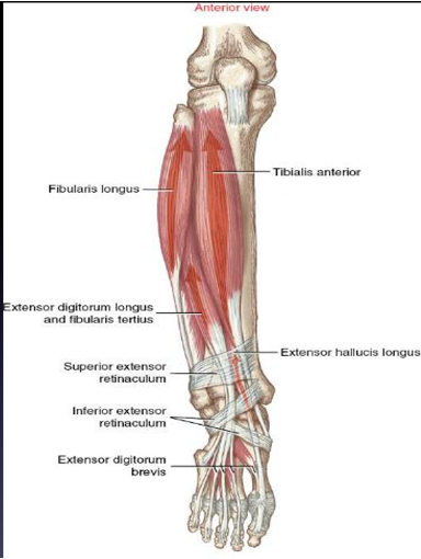
This takes us to the muscles that enable this movement. Recall that muscles can only pull. Thus, the tibialis anterior – we can feel this going over our shin bone – serves to lift our foot and toes skyward. This is called dorsiflexion – dorsi is Latin for top, in this case moving upward. Try to visualize the shin muscles (primarily the tibialis anterior) contracting and shortening. Do you see how the foot gets pulled upwards into dorsiflexion?
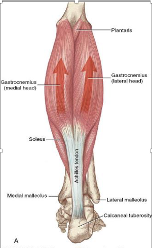
The calf muscles, our gastrocnemius and soleus, then pull to move the foot downward via the Achilles tendon. This is called plantarflexion – plantar meaning bottom.
Visualize those muscles contracting and shortening, pulling on the Achilles tendon, which then pulls the base of the heel upward, causing plantarflexion (like a ballerina pointing her toes).
Achilles Tendon
The Achilles is of particular importance to runners. It is the largest tendon in the body and serves as the springboard for upward and forward movement. It is named after the Greek hero Achilles, who was done in by an arrow piercing the back of his ankle, the weakest part of his body. While we may doubt the mythology, the fact is if our Achilles is out of commission so are we! We simply can’t move forward! The Achilles is subject to tremendous force, not just during running but anytime we bend our knee to pick something up. It connects to the calcaneus, our heel bone, by means of fibers that if overstretched strain or tear.
Foot strike
Assuming all our muscles, tendons, and ligaments are in good working order, then we meet the ground in various ways, depending on our natural gait. Many of us are heel strikers, some are toe strikers, while others, including me, meet the ground with our midfoot. There are convincing arguments for barefoot or minimalist shoe running, but it seems this works well for some runners but not others. There are also some brands with zero-drop soles, though they may still have a lot of cushion. And now we have the so-called Super Shoes, which have spurred performance in the top runners, but whose extra cushioning might be an issue for recreational runners if used frequently for training.
One-size-does-not-fit-all!
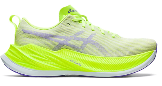
The reality is different runners have different needs when it comes to shoes and foot strike. The best advice I can offer here is to have several pairs of shoes you rotate so you have varying amounts of cushion and lift throughout your training.
Continuing with our gait, the normal foot meets the ground slightly on the outside part of the foot, then rolls inward – this is called pronation – and finally pushes off at the toes as our foot become air-bound. Pronation, per se, is not bad. It is a normal part of running gait.
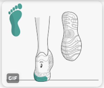
Yet, as with most things, there is a continuum, and many runners over-pronate which increases the amount of force that moves up the kinetic chain. Often the issues in our hips are really due to our feet!
Foot striking revisited
As noted above, there are three basic ways runners meet the ground. A debate has brewed for years about the best way for the foot to meet the ground. Heel strikers tend to hit the ground ahead of their center of mass, which results in a small amount of braking, a force we need to overcome and which puts stress up the kinetic chain. This tends to result in running inefficiency, since you are being pushed backwards slightly with each step.
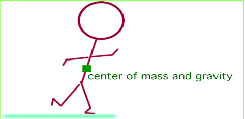
Forefoot and mid-foot strikers tend to hit the ground closer to their center of mass. While it might seem conclusive that heel striking should be avoided, many top runners find a heel-strike gait pattern works for them. So, the debate continues!
We’ve gone quickly through the lower extremity. We could easily have had a chapter on each segment. And maybe that will happen down the road. A key thing to keep in mind is as we age, injuries in our lower extremity, which I think we can concur will happen, simply take longer to heal. And that strength and balance become increasingly important with age.
Meanwhile, I hope what we’ve covered allows you to better understand how our running bodies function. The next two chapters will look at our various systems supporting running and health: the cardiovascular, respiratory, nervous, endocrine, and immune systems. We will then finish Part 1 with an chapter on nutrition. Part 2 will apply all that we have learned to our training and racing. I hope you are finding this series useful. Please subscribe if you haven’t already and send a link to those you think might find the content helpful.Take care, and keep running
